Ultrasound Scanning for Carcass Evaluation
Based on an article by Bruce Binnie, AgResearch, Ruakura
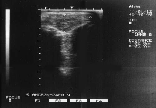
Ultrasound scanning can be a useful aid in assessing the breeding merit of animals within a flock for carcass composition. The estimation of eye muscle width, depth and fat in relation to the animal's live-weight can be used to predict the amount of lean meat and fat in the carcass.
Muscle Dimensions vs. Area
The same measurements can also be used to assess eye muscle area, but this does not necessarily mean a good estimation of weight of meat in the eye muscle. This is because scanning does not give an estimation of the length of the eye muscle, and so an animal may have a large eye muscle area, but it may also be shorter in the body, and therefore have no more total muscle weight. Indeed, large eye muscle area and shorter bodies often tend to go together.
Ruakura carcass data shows that eye muscle dimensions (width and depth) are more accurate predictors of composition (lean and fat) than eye muscle area (and eye muscle product). Further work is needed to find out whether the same applies to live animal scan data. There is some evidence that for eye muscle depth at least, scan data is more accurate than carcass data for predicting composition (Young, M. J. et al, 1996. Proceedings of the NZ Society of Animal Production. 56:205:211). So it is possible that scan eye muscle area may be a more accurate predictor of carcass lean and fat than scan eye muscle dimensions.
Similar approximations apply to estimations of eye muscle area from the product of width and depth. Different breeds, for example, are known to have different cross-sectional shapes of the eye muscle. Thus, different results will apply to two different breeds, and presumably to different animals within a breed. More work is needed to fine-tune these relationships.
Ranking Animals
Comparing the records of animals that have been born and reared in the same flock is the most usual way to use scan records. When there are genetic links between different flocks (e.g. through the use of reference sires), comparisons can also be made between flocks provided a good across-flock sire referencing analysis is carried out. IF THERE ARE NO GENETIC LINKS BETWEEN FLOCKS, ANIMALS CANNOT BE VALIDLY COMPARED ACROSS FLOCKS. This is especially so at shows and sales where each flock is usually represented by only one or a small number of animals. The different rearing circumstances found in the various flocks makes comparisons of animals from different flocks quite invalid.
Scanner Variations
For a within-flock comparison of breeding value, the aim is to rank the animals for lean and fat, and to assess the relative breeding merit of animals. It is possible to have two scanners who get different absolute values for eye muscle width, depth and fat, but who still do an adequate job of ranking the animals for breeding merit. Consider the following simple example:
| Animal | Scanner 1 | Scanner 2 | Scanners | |||
| A | B | A | B | A | B | |
| 1 | 60 | 32 | 65 | 34 | 54 | 29 |
| 2 | 50 | 26 | 55 | 28 | 45 | 23 |
| 3 | 56 | 28 | 61 | 30 | 50 | 25 |
| 4 | 58 | 28 | 63 | 30 | 52 | 25 |
| 5 | 54 | 26 | 59 | 28 | 49 | 23 |
| 6 | 54 | 28 | 59 | 30 | 49 | 25 |
When these data are put on a graph, it becomes clear that each operator has ranked the animals in the same order, with almost identical gaps between any two animals. In this case, the differences between operators is unimportant - they have all established the same relativities between the animals from the same flock. There should be real concern, however, when operators do not rank the animals in the same order, or they estimate markedly different gaps between animals.
Scanning accuracy for breeding value assessments depends on many factors, including:
- Rearing. The animals to be scanned should all have been born and reared on the same farm and under as near the same conditions as possible.
- Stance. The animals should be standing in a normal stance and be relaxed when the picture is frozen. The muscle shape changes significantly if the head is down or up, or if the animal is relaxed or tense.
- Site. The scanning site should be consistent. It is of less consequence whether the scanner elects to scan between the 12th and 13th ribs or behind the 13th rib provided every animal is scanned at the same site.
- Technique. The pressure of the scanning head against the skin should be consistent and as light as possible to maintain a picture. The fat layer, and to a lesser extent, the muscle shape can be distorted if the pressure of the head against the skin is too heavy. Avoiding variation of this pressure from one animal to another is most important for accurate relativity.
- Equipment. Like all machines, regular maintenance and calibration of the scanner's gear is important
- Interpretation. Consistent picture interpretation is probably where much variation between operators occurs. Some operators can capture a good picture, but fail to boundaries determining eye muscle width. Unless operators regularly have some stock scanned, slaughtered and measured, it is quite possible they will be unaware of this difficulty.
Scanning Site
There are two possible sites on the 12th or 13th ribs at which to scan - the "C" site or the "GR" site. The "C" site can be more accurate than the "GR" site because it is only fat, whereas the "GR" site is a tissue depth. However, there is less depth at the "C" site, and in very lean animals, where the fat depth may be only 2 or 3 mm, this can be a source of error. The ultrasound machine can be read to an accuracy of 1 mm. If the operator is 1 mm wrong, this is 33-50% wrong. The tissue depth at the "GR" site is about 2 or 3 times greater than at the "C" site, and so in the same very lean animals, a 1 mm error would be only a half or a third as great.
Most operators prefer scanning fat at the "C" site because they can do so on the same image as they do the eye muscle width and depth. In contrast, the operator has to capture a second image to get the "GR" site. So, if there is enough fat at the "C" site, it is the preferable option, both from an accuracy point of view, and also because the throughput of animals is faster.
There is no easy way to check the accuracy of a scanner who has been scanning at the "C" site. The carcasses have to be cut at the junction of the loin and rack, and an experienced technician then needs to measure the fat depth. Even then, the point is not whether the operator has measured the exact fat depth, but whether the operator has ranked the animals accurately.